Ultrasound of the Thyroid gland
 !
!
Introduction:
Thyroid nodule evaluation using ultrasound is presently viewed as a controversial topic. One of the main points of controversy on this topic is ultrasounds inability to accurately distinguish between a benign and malignant nodule just from images alone
Controversy is also linked to the detection rates of thyroid nodules which have been steadily increasing over the past years although there has not been a matching increase in mortality rates. The literature states that this could be due to multiple factors like ultrasound technology becoming more sensitive thus detecting more nodules
Body of Review:
Sonographic features and their different appearances between a benign and malignant nodule will be discussed using the latest guidelines from the American Thyroid Association (ATA). The role that ultrasound has played and currently is playing in the assessment of these nodules will be discussed as well as new techniques and technologies becoming available. It is hoped that these new techniques and technologies could aid Sonographers and Radiologists to better distinguish between a benign and malignant nodule, promoting an improved selection criterion for when fine needle aspiration (FNA) of the nodule should be utilised and lower the current controversy

Thyroid gland:
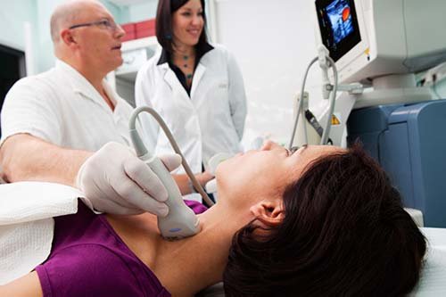
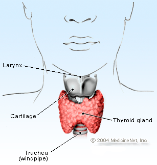
Ultrasound can study the thyroid gland in detail due to the glands superficial location in the anterior inferior region of the neck. The thyroid is butterfly shaped gland consisting of 2 lobes. The function of the gland is to provide the body with adequate amounts of thyroxine (T4), triiodothyronine (T3) and calcitonin hormones. The T4 and T3 hormones are stored in colloid within the follicles of the gland which are the functional units. An ultrasound thyroid examination consists of assessing the echogenicity, echo texture, vascularity, total volume size of the bilateral lobes and isthmus. Pathology detected should be measure in 3 plains and its vascularity assessed
Thyroid Nodules:
The terminology of the word “thyroid nodule” is used in the literature as a broad word to describe any growth of cells that has formed a lump or mass located on the gland itself. These nodules could be simple colloid cysts, soil nodules consisting of normal thyroid parenchyma, or focal nodules consisting of different types of other thyroid parenchyma. Thyroid nodules are not described as one single pathology but are described as a wide range of different Pathologies.
Thyroid nodules can either be benign or malignant, studies have shown that 95% of these detected nodules are in fact benign. These benign nodules can either be colloid nodules (cysts) or follicular benign nodules which are soil growths. If a nodule is found to be malignant 75% of the time the malignant nodule is found to be a papillary carcinoma making it the most common type of thyroid cancer.
The Role of Ultrasound in Detecting Thyroid Nodules:
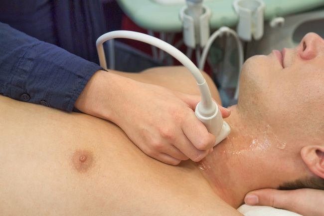
Presently linear array ultrasound transducers can operate at up to a frequency of 15 MHz and to a depth of 5cm with a resolution of 0.5 to 1.0 mm. No other medical imaging method can achieve this level of spatial resolution (ability to differentiate between two separate echoes). The thyroid gland is a superficial gland therefore ultrasound plays an important role in helping diagnose and evaluate detected thyroid nodules.

Currently when using ultrasound there is no single suspicious sonographic feature that can accurately distinguish or is sensitive enough to diagnose a thyroid nodule as being malignant. Ultrasound cannot give a 100% correct diagnoses that determines whether a nodule is either malignant or benign it can only can only speculate the probability which is where much of the controversy lies and challenges radiologists.
Fine Needle Aspiration:
The next step for a detected nodule that has suspicious sonographic features is fine needle aspiration (FNA) to gain an accurate diagnosis. FNA allows a pathologist to sample and assess the suspicious nodules cellular parenchyma under microscope that would be impossible to assess just by ultrasound. Fine need aspiration (FNA) was first introduced in the late 1970s with great success and has been responsible for halving the number of patients that would have otherwise received a thyroidectomy because there was no way of diagnosing if the detected nodule was malignant or not thus savings. FNA has become and still is the primary method for diagnosing malignant nodules which has the highest specificity rate of 72 – 100%.
In the past FNA of nodules did have its limitations in diagnosing nodules. These limitations noted were due to lack of operator expertise such as not sampling enough of the cellular parenchyma, contaminating the sampling smears, error in patient’s profile, and sampling normal or the wrong cellular parenchyma from the suspicious nodule therefore creating high false negative results.
Now with the establishment of ultrasound guided fine needle aspiration (UGFNA) a lot of the previous limitations have now been eliminated due to a more precise needle tip placement within the nodule. UGFNA is now the primary method of evaluating suspicious thyroid nodules and in 2011 FNA by means of simple palpation accounted for just 6.2% of procedures while ultrasound UGFNA accounted for 93.8%. UGFNA has led to a 3 to 5-fold increase in satisfactory cellular sample rates than compared to conventional palpation FNA and the false negative rates are at 9.1%.
Increase in the Detection of Thyroid Nodules
Data from America has found that since 1975 the incidences of detected nodules increased by from 4.9 to 14.3 per 100,000 individuals and much of the other literature is consistent with this increase. It is suggested that using ultrasound to evaluate the thyroid gland against the traditional palpation methods has been largely responsible for the increase of incidental findings of nodules. This suggestion has been supported by the fact that 87% of these incidence findings are from nodules that are < 2cm in diameter which can now be detected by ultrasound.
Advances in Ultrasound Technology:
Over the decades marked advancement in ultrasound technology has taken place. These advancements such as harmonic imaging, compound imaging and improved spatial resolution have all contributed to the increase of incidental detection rates of thyroid nodules. Modern day ultrasound transducers can now operate at higher frequencies due to the increased number of Lead Zirconate Titanate (PZT) elements packed into the head of the transducer resulting in increased bandwidths that were never before possible by previous transducers.
Ultrasound contrast resolution refers to the machines ability to distinguish between echoes of different amplitudes which originate from adjacent and nearby structures. Improvements in contrast resolution has been found as being one of the primary factors that have influenced the increased detection of thyroid nodules and the incidental detections of these nodules
Machine echo processing techniques have also been improved and now use a system known as speckle reduction (signal processing). Speckle reduction works by better integrating the pixels related to the received echoes on to the monitor screen which improves the signal to noise ratio by using pixel screen recognition. For example, when an image of a colloid cystic nodule is being is evaluated with ill-defined margins and prominent artefacts using speckle reduction it is now possible for the unwanted artefacts and ill-defined margins to be removed and the entire image is cleaned up and made to appear much sharper and recognized as a true colloid cyst.
Future Technology:
Even though there has been continued improvements in ultrasound technology empirical evidence suggests that ultrasound still cannot distinguish between a benign and malignant nodule. Wenzhou central hospital located in China recognised the importance of being able to distinguish between a benign and malignant nodule. Currently a radiologist makes the diagnoses but this method is not always accurate and errors can occur. Computer aided diagnostic systems (CAD) uses pattern recognition for recognising malignant nodules and is used to aid radiologists. More recently Wenzhou central hospital has developed a more advanced CAD known as an extreme learning machine (ELM). This system was programed with the 5 sonographic features associated with nodule malignancy, Hypoechoic, Internal Micro Calcifications, Irregular margins, Taller than wider in shape, Rim calcification. Out of the 187 patents with confirmed malignant nodules the ELM system could detect between 90.6% and 98% of the malignant nodules.
Hard thyroid nodules are suspicious for cancer so a technique known as ultrasound Elastography (USE) was developed to assess the stiffness of these suspicious nodules. By applying external physical pressures an assessment of the compressibility and strain that occurs within the nodule can be made. This technique requires high operator skills and can only produce semi quantitative images and is not capable of precisely measuring velocities, at best it is 71% accurate (Baskin, 2013, p.355). Shear wave Elastography (SWE) is an even newer technique which uses acoustic impulses to stimulate tissues and generate localized lateral displacements called “shear waves” and is used in conjunction with conventional 2d, at best it is 92% accurate and imaging is operator independent and is more sensitive than USE.
Summary:
The literature states that 95% of nodules are benign with the other 5% being malignant. If nodules are benign then they can present as simple colloid cysts, soil nodules of normal thyroid parenchyma, or a nodule consisting of different types of other thyroid parenchyma. If nodules are found to be malignant then 75% of the time that nodule will be diagnosed as a being a papillary carcinoma.
Palpation of the gland by a physician can detect only 4 - 5% of thyroid nodules. However, when ultrasound is utilized the detection rate of nodules increases by 50% providing evidence that ultrasound is a superior method of imaging for detecting nodules over any other imagery.
Sonographers using ultrasound can only detect and evaluate the sonographic features of detected nodules. Ultrasound cannot diagnose a nodule from being malignant or benign as there is no single suspicious sonographic feature this can only be diagnosed by FNA.
Ultrasound becomes more effective when used in conjunction with the updated ATA guidelines as it gives Sonographers and Radiologist’s better guidance as to what constitutes as malignant and benign. But again the ATA at its best can give a 90% probability that a nodule is malignant if it matches the “highly suspicious” category.
Conventional FNA has been replaced by UGFNA due to its high false negative rates. UGFNA has a 3 to 5-fold increase in satisfactory cellular sample rates and a 9.1% false negative rate.
UGFNA is still the primary method for diagnosing malignant nodules with a specificity rate of 72 – 100% accuracy.
USE is 71% accurate in diagnosing malignant nodules and SWE is 92% accurate. It appears SWE is such a new technique that more studies are needed to gain a better conclusion.
ELM was shown to have detected 90.6% and 98% of the malignant nodules out of 187 patents with confirmed malignant nodules. Although this is still a very new technology it has shown already impressive results and continued improvements could raise its detection rate higher.
Conclusion and Critical Analysis:
Technology improvements has helped ultrasound become more accurate in detecting nodules but Sonographers and Radiologists can only speculate the probability of a nodule being malignant based on its current sonographic features.
The updated ATA guidelines is helping Sonographers and Radiologists determine what constitutes as a malignant and benign nodule but still UGFNA is the only way to diagnose a malignant nodule. It is highly likely that this controversy will continue into the future as it is impractical to biopsy every single patient presenting with a highly suspicious nodule.
Even though UGFNA has greatly improved the accuracy of diagnosis errors can still occur and this appears to be due to operator error, an increase in training and experience may solve these problems.
Ultrasound can now detect nodules < 2cm in diameter and it is highly probable that these advancements in ultrasound technology have contributed to an increase in the number of nodules being detected. Also there has been no increase in mortality rates associated with the increase in these detection rates. There has been an increase in nodules but it seems rather there has been an epidemic of more nodules now being detected.
With ELM shown to have diagnosed malignant nodules at a rate of 90.6% and 98% of the time it has already shown great progress and may end the controversy in the future even making biopsies obsolete.
Very scary that thyroid cancer has trippled in the last couple decades. Is it Fukushima? Auto-immune on the rise from GMO food? Increase in cancer therefore cancer treatment (especially radiation absorbed by the thyroid)? Increased technology should theoretically detect more nodules, but to get that test ordered by a doctor in Canada is not easy - you practically have to have a giant goitre sticking out of your neck.
Medikatie. Thank you for your reply. I dont think that thyroid nodules have increased over the decades. More the fact that ultrasound technology has improved so much that it is now detecting more thyroid nodules. The thyroid nodules have always been there, just that now they are being detected with improved ultrasound technology.