UPDATE FROM SFHEALTH
The muscular system is an organ system consisting of skeletal, smooth and cardiac muscle. It permits movement of the body, maintans posture and circulate blood throughout the body. But the skeletal muscles and its deseases described in this post are those not directly involved in the movement of the joints of the limbs.
⬛MUSCLE OF THE FACE

There are a large number of muscle involved in changing facial expression and with movement of the lower jaw during chewing and speaking. This muscles are: occipitofrontalis, levator palpebrae superioris, orbicularis, buccinator, orbicularis Oris, masseter, temporalis, pterygoid muscle.
▪️OCCIPITOFRONTALIS (UNPAIRED): Consist of a posterior muscular part over the occipital bone, an anterior part over the frontal bone and an extensive flat tendon or aponeurosis that stretches over the done of the skull and joints the two muscular parts. It raises the eyebrows.
▪️LEVATOR PALPEBRAE SUPERIORIS: Extends from the posterior part of the orbital cavity to the upper eyelid. It raises the eyelid.
▪️BUCCINATOR: This flat muscle of the cheek draws the cheeks in towards the teeth in chewing and in forcible expulsion of air from the mouth.
▪️ORBICULARIS ORIS (UNPAIRED): Sorounds the mouth and blends with the muscles of the cheek. It closes the lips and, when strongly contracted, shapes the mouth for whistling.
▪️MASSETER: Is a broad muscle, extending from the zyvomatic arch to the angle of the jaw. In chewing it draws the mandible up to the maxilla and exerts considerable pressure on the food.
▪️TEMPORALIS: Covers the squamous part of the temporal bone. It passes behind the zyvomatic arch to be inserted into the coronoid process of the mandible. It closes the mouth and assist with chewing.
▪️PTERYGOID MUSCLE: Extends from the sphenoid bone to the mandible. It closes the mouth and pulls the lower jaw.
⬛ MUSCLES OF THE NECK

There are a great many muscles situated in the neck but the largest two are; sterbocleidomastoid muscle and trapezius muscle.
▪️STERBOCLEIDOMASTOID MUSCLE: Arise from the manubrium of the sternum and the clavicle and exstends upwards to the mastoid process of the temporal bone. It assists in turning the hand from side to side. When the muscle on one side contract together they flex the clavicle upwards when the head is maintained in a fixed position, e.g., in forced respiration.
▪️TRAPEZIUS MUSCLE: Covers the shoulder and the back of the neck. The upper attachment is to the occipital protuberance; the medial attachment is to the transverse processes of the cervical and thoracic vertebrae; and the lateral attachment is to the clavicle and to the spinous and acromion processes of the scapula. It pulls the head backwards, squares the shoulder and controls the movements of the scapula when the shoulder joints is in used.
⬛ MUSCLE OF THE BACK
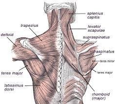
There are six pairs of large muscles in the back in addition to those that form the posterior abdominal wall. The arrangement of this muscles is the same on each side of the vertebral column. They are: trapezius, teres major, psoas, quadratus lumborum, sacrospinalis, latissimus dorsi.
▪️QUADRATUS LUMBORUM: Originates from the crest of the illium then it passes upwards, parallel and close to the vertebral column and is inserted into the 12th rib. Together the two muscles fix the lower rib during respiration and cause extension of the vertebral column. If one muscle contracts it causes flexion of the lumbar region of the vertebral column.
▪️SACROSPINALIS: Is a group of muscles, lying between the spinous and traverse processes of the vertebrea. They originates from the sacrum and are finally inserted into the occipital none. Their contraction causes extension of the vertebral column.
⬛ MUSCLES OF THE ABDOMINAL WALL
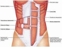
◾ANTERIOR AND LATERAL PARTS: There are four pairs of muscles, arranged in four layers, that make up the anterior and lateral parts of the abdominal wall. From the surface inwards they are: rectus abdominis, external oblique, internal oblique, internal oblique, transversus abdominis.
The anterior abdominal wall is divided longitudinally by a very strong midline tendinous cord, the linea alba,
which extends from the xiphoid process of the sternum to the symphasis pubis. The structure of the abdominal wall on each side of the linea alba is identical.
▪️RECTUS ABDOMINIS: Is the most superficial muscle. It is broad and flat, originating from the transverse part of the public bone then passing upwards to be inserted into the lower rib and the xiphoid process of the sternum. Midially the two muscles are attached to the Linea alba.
▪️EXTERNAL OBLIQUE: Exstends from the lower ribs downwards and forward to be inserted into the illiac crest and by an aponeurosis, to the Linea alba.
▪️INTERNAL OBLIQUE: Lies deep to the external oblique. It fibers arise from the crest of the illium and by a broad band of facia from the spinous processes of the lumber vertebrea. The fibers pass upwards towards the midline to be inserted into the lower ribs and by an sponeurosis, into the Linea alba. The fibers are at right angles to those of the external oblique.
▪️TRANSVERSUS ABDOMINIS: Is the deepest muscle of the abdominal wall. The fibers arise from the illiac crest and the lumber vertebrea and pass across the abdominal wall to be inserted into the Linea alba by an aponeurosis. The fibers are at right angles to those of the rectus abdominis.
◾POSTERIOR ABDOMINAL WALL: The muscles of the posterior part of the abdominal wall are: quadratus lumborum, psoas, internal oblique, transversus abdominis.
⬛ MUSCLES OF THE PELVIC FLOOR

The pelvic floor is divided into two identical parts at the midline. Each half is made up of muscles and fascia which unite in the midline. The muscles are: levator Ani and cocygeus.
▪️LEVATOR ANI: Are broad flat muscles, forming the anterior part of the pelvic floor. They originate from the inner surface of the true pelvis and unite in the midline. Together they form a slug which supports the pelvic organs.
▪️COCYGEUS: Cocygeus are triangular sheets of muscle and trendinous fibers situated behind the levator ani. They originate from the medial surface of the ischium and are inserted into the sacrum and coccyx. They complete the formation of the pelvic floor which is perforated in the male by the urethra and anus, and in the female by the urethra, vagina.
⬛ MUSCLE OF RESPIRATION
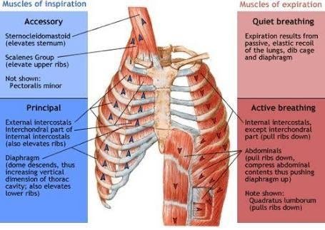
The muscles of respiration are: External intercostal, internal intercostal, diaphragm, where external intercostal has 11 pairs, Internal intercostal has 11 pairs same as the external intercostal. While the diaphragm has only 1 pair.
⛑️ DISEASES OF MUSCLES
⚫MYASTHENIA GRAVIS
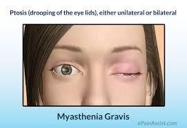
This is an autoimmune disease of unknown origin in which there is defective muscle stimulation. Antibodies develop which damage the acetylcholine receptors of neuromuscular junctions, by blocking the transmission of the nerve impulse to muscle fibers. This causes progressive and and extensive muscle weakness. Extraocular and eyelid muscles are affected first, followed by those of the neck and limbs. There are periods of remission, relapses being precipitated by, e.g., severe muscular exercise, infections, emotional disturbances, pregnancy.
⚫MHOPATHIES:

▪️PROVRESSIVE MUSCULAR DYSTROPHIES: In this group of inherited disease there is progressive degeneration of groups of muscles. The main differences in the types are the:
Age of onset
Rate of progression
Groups of muscles involved
▪️DUCHENNE TYPE: The muscles abnormality is present before birth but may not be evident untill the child is about 5 years of age. Wasting and weakness begin in muscles of the lower limbs then spread to the upper limbs, progressing without remission. Death usually occurs in adolescence, often from infection.
▪️FACIO-SCAPULO-HUMERAL DYSTROPHY: This disease usually begins in the adolescence and the younger the age of onset the more rapidly it progresses. Muscles of the face and upper limbs are affected first. This is a chronic condition that usually progresses slowly and may not cause complete disability.
▪️MYOTONIC DYSTROPHY: This disease usually begins in adult life. Muscles contract and relax slowly, often seen as difficulty in releasing an object held in the hand. Muscles of the tongue and the face are affected then muscles of the limbs. Other conditions associated with myotonic dystrophy include:
Premature cataract
Atrophy of the gonads
Defects in the conducting system of the heart
Endocrine disturbances, e.g., in insulin utilization the disease progresses without remission and with increasing disability. Death usually occurs i in the middle age.

REFERENCES
THANK YOU FOR READING, HOPE YOU'VE LEARNED SOMETHING TOO I KNOW I DID
TILL NEXT TIME.

Lovely written friend
You're making me like biology...
Lovely written bro. Keep up the good work.
Am happy to read this, you refresh my memory.
Thank you @ninoh22
Well detailed..... Good one @fidelmboro
Great one @fidelmboro
You gave a very detailed analysis to this...
flagged for plagiarism @steemflagrewards
Source
Steem Flag Rewards mention comment has been approved! Thank you for reporting this abuse, @mathowl categorized as plagiarism. This post was submitted via our Discord Community channel. Check us out on the following link!
SFR Discord
This is very informative. Well done mate!
Where do you get this definition? Is muscular system really an organ system?
It's difficult for a layman to comprehend a medical-related article especially when it concerned with the anatomy. I guess, it's worth trying but if there were too much unfamiliar words, it can be impossible to be read by a layman.
It's straight copied from the Wikipedia article.
But don't bother expecting too much from this author - most content of this post is plagiarized anyway.
I see. Too bad for the author then.
:)))
Great enlightenment there.
Well done