Hello steemians ,
This is my first post related to science.
I'm here to explain the interesting topic, heart.May the first post be the best post.
Introduction:-
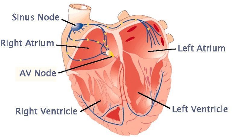
Heart is a powerful muscle that pumps blood throughout the body by means of a coordinated contraction.The contraction is generated by an electrical activation,which is spread by a wave of bioelectricity that propagates in a coordinated manner throughout the heart.Under normal conditions,the sinoatrial node initiates an electrical impulse that propagates through the atria to the atrioventricular node,where a delay permits ventricular filling before the electrical impulse proceeds through the specialized His-Purkinje conduction system that spreads the electrical signal at speeds of meters per second throughout the ventricles.
This electrical impulse propagates diffusively through the heart and elevates the voltage at each cell, producing an action potential,during which a surge in intracellular calcium initiates the mechanical contraction.The normal tachycardia and fibrillation.
Heart Anatomy and Structure:-
Heart:
A powerful muscle slightly larger than a clenched fist.It is composed of four chambers, two upper(artia) and two lower(ventricles).It works as pump to send oxygen-rich blood through all the parts of the body.A human heart beats an average of 100,000 times per day.During that time, it pumps more than 4,300 gallons of blood throughout the entire body.
Right ventricle:
The lower right chamber of the heart. During the normal cardiac cycle, the right ventricle receives deoxygenated blood as the right atrium contracts.During this process the pulmonary valve is closed, allowing the right ventricle to fill.Once both ventricles are full, they contract. As the right ventricle contracts, the tricuspid valve closes and the pulmonary valve opens. The closure of the tricuspid valve prevents blood from returning to the right atrium, and the opening of the pulmonary valve allows the blood to flow into the pulmonary artery toward the lungs for oxygenation of the blood.
The right and left ventricles contract simultaneously;however,because the right ventricle is thinner than the left, it produces a lower pressure than the left when contracting.This lower pressure is sufficient to pump the deoxygenated blood the short distance to the lungs.
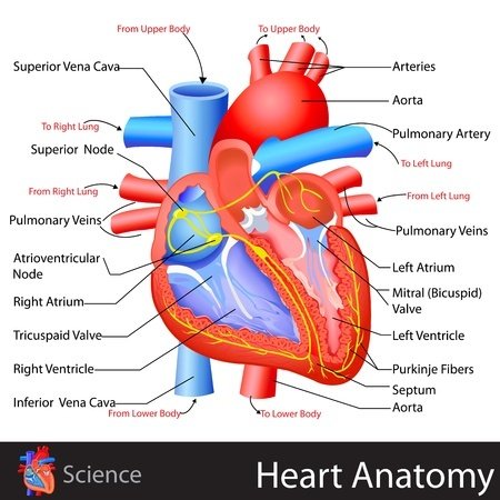
Left ventricle:
The lower left chamber of the heart. During the normal cardiac cycle, the left ventricle recieves oxygenated blood through the mitral valve from the left atrium as it contracts. At the same time, the aortic valve leading to the aorta is closed, allowing the ventricle to fill with blood. Once both ventricles are full, they contract. As the left ventricle contracts, the mitral valve closes and the aortic valve opens. The closure of the mitral valve prevents blood from returning to the left atrium, and the opening of the aortic valve allows the blood to flow into the aorta and from there throughout the body. The left and right ventricles contract simultaneously; however because the left ventricle is thicker than the right, it produces a higher pressure than the right when contracting. The higher pressure is neccessary to pump the oxygenated blood throughout the body.
Right Atrium:
The upper right chamber of the heart. During the normal cardiac cycle, the right atrium receives deoxygenated blood from the body. Once both atria are full, they contract, and the deoxygenated blood from the right atrium flows into the right ventricle through the open tricuspid valve.
Left Atrium:
The upper left chamber of the heart. During the normal cardiac cycle, the left atrium receives oxygenated blood from lungs through the pulmonary veins. Once both atria are full, they contract, and the oxygenated blood from the left atrium flows into the left ventricle through the open mitral valve.
Superior Venacava:
One of the two main veins bringing deoxygenated blood from the body to the heart. Veins from the head and upper body feed into the superior venacava, which empties into the right atrium of the heart.
Interior Venacava:
One of the two main veins bringing deoxygenated blood from the body to the heart. Veins from the legs and lower torso feed into the interior vena cava,which empties into the right atrium of the heart.
Aorta:
The central conduit from the heart to the body, the aorta carries oxygenated blood from the left ventricle to various parts of the body as left ventricle contracts. Because of the large pressure produced by the left ventricle, the aorta is the largest single blood vessel in the body and is approximately the diameter of the thumb.
Atrial septum:
The wall between the two upper chambers both the right and left atrium of the heart.
Pulmonary trunk:
A vessel that conveys deoxygenated blood from the right ventricle of the heart to right and left pulmonary arteries, which procceed to lungs.
Pulmonary veins:
The vessels that transport oxygenated blood from the lungs to the left atrium. The pulmonary veins are the only veins to carry oxygenated blood.
Pulmonary Valve:
One of the four one-way valves that keep blood moving properly through the various chambers of the heart+ The pulmonary valve separates the right ventricle from the pulmonary artery.
Aortic valve:
One of the four one-way valves that keep blood moving properly through the various chambers of the heart. The aortic valve, also called a semi-lunar valve, seperates the left ventricle from the aorta.
Mitral valve:
One of the four one-way valves that keep blood moving properly through the various chambers of the heart. Located between the right atrium and the right ventricle, the tricuspid valve is the first valve that blood encounters as it enters the heart. When open, it allows the deoxygenated blood collected in the right atrium to flow into the right ventricle.
Atria:
The two upper cardiac chambers that collect the blood entering the heart and send it to the ventricles. The right atrium receives blood from the superior vena cava and interior vena cava. The left atrium receives blood from the pulmonary veins.
Ventricles:
The two lower cardiac chambers that collect blood from the upper chambers and pump it out of the heart. Because the ventricles pump blood away from the heart, they have thicker wall than the atria so that they can withstand the associated higher blood pressures.
Blood Flow:
The heart's cycle begins when oxygen-poor blood from the body flows into the right atrium. Next the blood flows through the right atrium into the right ventricle, which serves as a pump that sends the blood to the lungs. Within the lungs, the blood releases waste gases and picks up oxygen. This newly oxygen-rich blood returns from the lungs to the left atrium through the pulmonary veins. Then the blood flows through the left atrium into the left ventricle. Finally, the left ventricle pumps the oxygen-rich blood out through the aorta and from there to all parts of the body. The human body has about 5.6 liters of blood, all of which circulates through the body three times every minute.
.gif)
Arrhythmias:
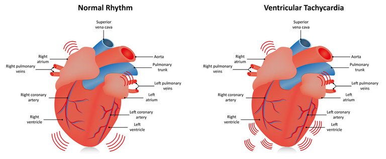
During normal rhythm, the heart beats regularly, producing a single coordinated electrical wave that can be seen as a normal electrocardiogram(ECG). During arrhythmias such as ventricular tachycardia and ventricular fibrillation, this normal behaviour is disrupted and the ECG records rapid rates with increased complexity.
The underlying cause of many arrhytmias is the development of a reentrant circuit of electrical activity that repetitively stimulates the heart and produces contractions at a rapid rate. During tachycardia, a single wave can rotate as a spiral wave,producing fast rates and complexity. During fibrillation, a single spiral wave can degenerate into multiple waves. Because contraction is stimulated by the pattern of electrical waves, arrhythmias can compromise the heart's ability to pump the blood and sometimes may be lethal.
Sinus rhythm:
This is the normal rhythm of the heart and results from the proper activation of the entire heart in proper sequence. The sinus located in the right atrium spontaneously beats periodically to begin each activation, which proceeds to activate both atria before reaching the atrioventricular(AV) node. At the AV node, which under normal circumstances is the only electrical connection between the atria and ventricles, activation proceeds more slowly to allow for optimal ventricular filling. The activation then enters the His-Purkinje system, which allows a rapid spreading throughout the ventricles and induces a strong, synchronized contraction. Any variation from normal sinus rhythm is termed an arrhytmia.
Ventricular fibrillation:
This is an immediately life-threatening arrhythmia in which the heart's electrical activity and associated contraction become discorded and ineffective. It is characterized by rapid, irregular activation of the ventricles and thereby prevents an effective mechanical contraction. Blood pressure instantaneously drops to zero,leading to death within minutes due to lack of cardiac output unless successful electrical defibrillation is performed; spontaneous conversion to sinus rhythm is rare. During ventricular fibrillation, the ECG has no distinctive QRS complexes but instead consists of an undulating baseline of variable amplitude. Although the sinus node continues to function properly, P waves cannot be discerned in the VF waveform. Ventricular tachycardia in many cases can degenerate to ventricular fibrillation. One mechanism believed to be responsible for rapid, unsynchronized contraction of the ventricles is the continuos reactivation of one or more electrical circuits in the ventricles, which can appear as three- dimensional spiral or scroll waves of electrical activity. These waves prevent the ventricles from contracting in a coordinated manner, thereby compromising the heart's ability to pump blood.
Ventricular tachycardia:
This is a rhythm characterized by wide, bizzare QRS complexes and frequent ventricular premature contractions in a row. VT may be paroxysmal or chronic and often signifies underlying myocardial disease. Mechanisms include reentry, increased automatically, and trigged activity. VTis potentially life-threatening, as it can degenerate rapidly to ventricular fibrillation and result in sudden death.
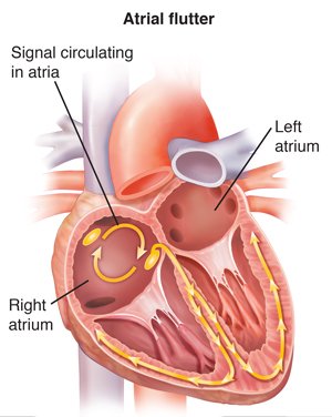
Artial flutter:
This is an AV node-independent intra-artial macro-reentry rhythm, in which the atrial anatomy sustains a loop of continuous depolarization, often around the tricuspid valve annulus in the right atrium. Atrial flutter can be paroxysmal or chronic and may be associated with extremely rapid ventricular response rates. variable degrees of AV block often modify AV conduction, which changes between 1:1 and 3:1 or 2:1. Atrial flutter often induces electrical remodeling and thereby can serve as a precursor to atrial fibrillation.
Atrial fibrillation:
This is characterized by rapid, irregular activation of the atria. Causes can include reentry and abnormal automatically. Atrial fibrillation is one of the most commonly seen supraventricular arrhythmias.
AF can be paroxysmal or chronic. Chronic AF often causes a markedly elevated ventricular response rate and thereby contributes to the clinical signs of heart failure. AF also can occur in the absence of overt structural heart disease as lone AF, which can feature a normal.
.jpg)

Hi! I am a robot. I just upvoted you! I found similar content that readers might be interested in:
https://www.scribd.com/document/48637990/Heart-3d