What is MRI?
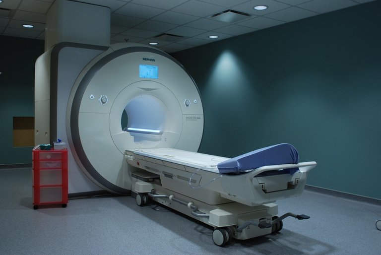
Magnetic Resonance Imaging is a scanning technique which utilizes strong magnetic fields and radio frequency waves to create extremely detailed images of the human body. It involves the use of a MRI machine - the large, doughnut-shaped machine in the image below. MRI machines have strengths of up to 7 tesla - about up to 7000 times the strength of a refrigerator magnet.

During a MRI scan, the patient is kept completely still through the use of straps - even the smallest movement will cause artifacts and blurring in the final image.
How does MRI work?
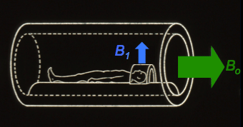
Hydrogen is a plentiful atom in the human body, in the form of H2O. The most abundant isotope of hydrogen, protium, only has a single proton in its nucleus. Protons have a positive charge - and as such, their magnetic moments (or spins) will align with the magnetic field generated by the MRI machine.
A radiofrequency pulse is then emitted by the MRI machine - a short rotating magnetic field generated in a perpendicular fashion to the main, static magnetic field generated by the main superconducting magnet.

This causes the protons to align to the new magnetic field briefly. While the protons are brought back to their equilibrium positions, they perform a rotating motion which induces a voltage in the receiver coil. Using something called a magnetic field gradient, the MRI machine can localize the image - choosing a specific area to image.
The current generated in the receiver coil is then parsed by computer systems to generate an image. Because the image is generated from the presence of hydrogen in body tissues, MRIs basically image the location of water and fat.
How do MRIs differentiate between types of tissue - say, between tumor tissue and healthy human tissue?
Protons in different tissues have different rates of returning to their equilibrium position - which means that healthy brain tissue would have a different signal to brain tumor tissue.
As well as this, before taking an MRI, sometimes patients are made to ingest a contrast agent - a chemical that accumulates in the target tissue and shows up very clearly on a MRI scan. This is the norm on other types of diagnostic imaging. A PET scan, for example, requires the intake of a barium meal which increases contrast in certain tissues.
Note to Readers
If there are any corrections, pieces of feedback or any suggestions for future posts you would like to put forward, please do put them down in the replies section.
If you would like to get informative tidbits of knowledge in your feed, please consider following me. It means a lot, and means that my content reaches more people.
Please do resteem and upvote this post if you found it interesting!
Thanks, Jian.
Heya

Hello, interestingly written, good luck! Come to my blog
Thanks guys! A follow would be much appreciated if you like my content :)
Congratulations @jiansoo! You received a personal award!
Click here to view your Board of Honor
Do not miss the last post from @steemitboard:
Congratulations @jiansoo! You received a personal award!
You can view your badges on your Steem Board and compare to others on the Steem Ranking
Vote for @Steemitboard as a witness to get one more award and increased upvotes!