Hello everyone,how are you all? Hope you all are doing well.we have learnt so much from abdomen anatomy.now we are going to learn about the last one which is inferior vena cava and kidney.
INFERIOR VENA CAVA
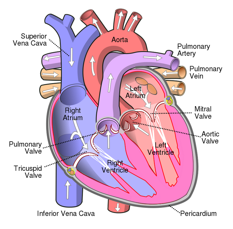
The inferior vena cava formed as the left and right common iliac veins behind the abdomen unite, at about the level of L5. It passes through the thoracic diaphragm at the caval opening at the level of T8.It passes to the right of the descending aorta.after passing through the venacaval opening it then enters into right atrium of heart.the inferior venacava carries the deoxygenated blood from the lower and middle body into the right atrium of the heart.
Below image is the inferior venacava in the abdomen
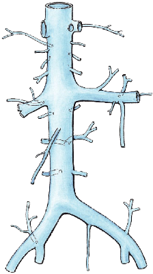
Image source
You can see in the below image the inferior vena cava draining into rigjt atrium.
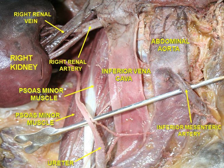
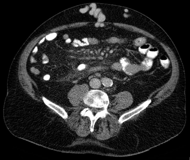
Beloe image is cadaveric image of inferior venacava and you can see abdominal aorta beside to it.you can notice the inferior vena cava is thin and abdominal aorta is thicker.
Tributaries of inferior vena cava
1)Hepatic veins
2)Inferior phrenic vein
3)Right suprarenal vein
4)Right renal vein
5)Right gonadal vein
6)L1-L5 lumbar veins
7)Right and left common iliac veins
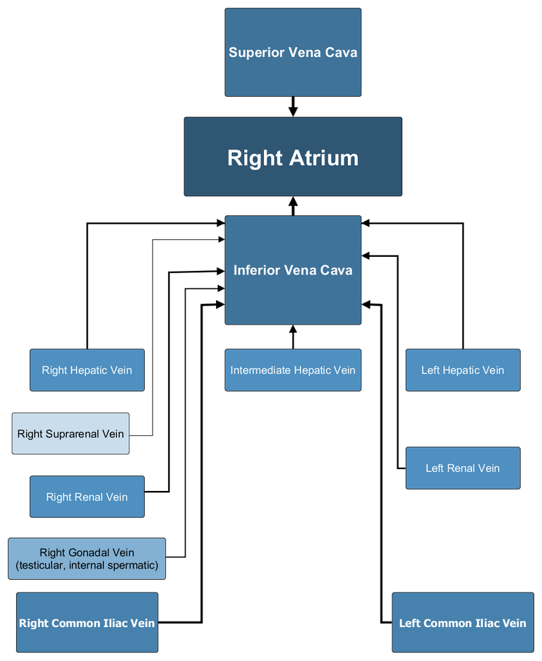
On the right side, the gonadal veins and suprarenal veins drain into the inferior vena cava directly.but on the left side, they drain into the left renal vein which in turn drains into the inferior vena cava.
You can understand tributaries by below image
Portocaval anastomosis
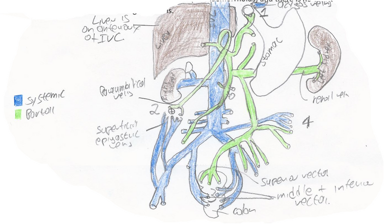
A portacaval anastomosis or portocaval anastomosis is a specific type of circulatory anastomosis that occurs between the veins of the portal circulation and the vena cava,thus forming one of the principal types of portosystemic anastomosis or portosystemic anastomosis, as it connects the portal circulation to the systemic circulation, providing an alternative pathway for the blood.
See the below image for better understanding
Clinical significance
In portal hypertension, as in the case of cirrhosis of the liver, the anastomoses become congested and form venous dilatations. Such dilatation can lead to esophageal varices and anorectal varices and Caput medusae can result.
| Sites | portal system | systemic system | name of clinical condition |
|---|---|---|---|
| Rectum | superior rectal vein | middle and inferior rectal vein | Haemorrhoids |
| Oesophagus | oesophageal branch of left gastric vein | oespphageal branch of accessory hemiazygous vein | Oesophageal varices |
| Umbilicus | paraumbilical vein | superficial veins of anterior abdominal vein | Caput medusae |
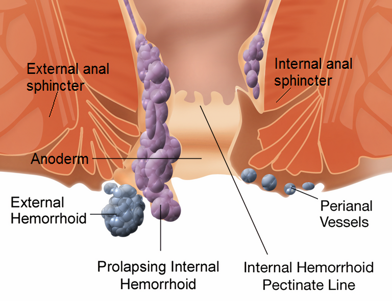
You can see below image of clinical conditions
Oesophageal varices
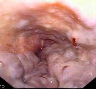
Image source
Haemorrhoids
Caput medusae
KIDNEY
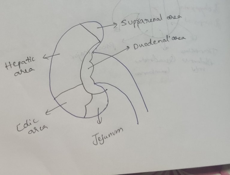
There are two kidneys in our body.we learnt the blood supply of kidney.now we are going to learn about relations of kidney
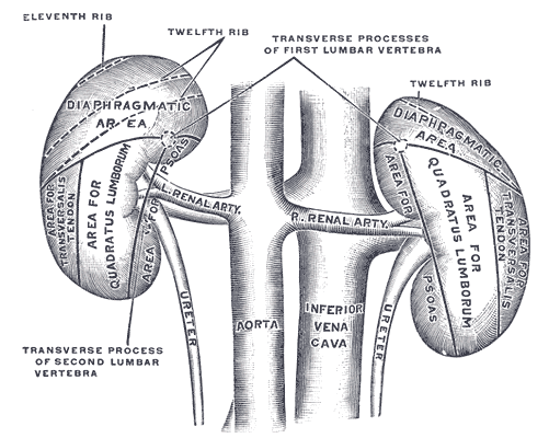
Posterior relations of kidney
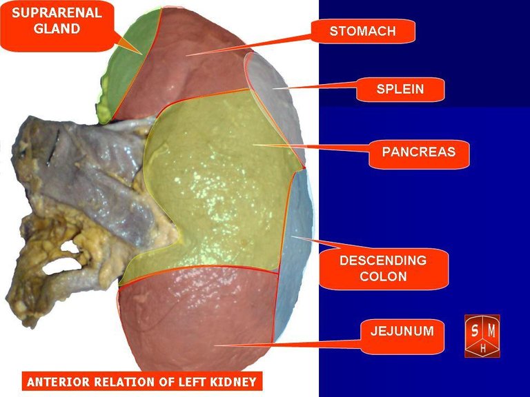
Anterior relations of left kidney
Anterior relations of right kidney
Below i have drawn a schematic diagram.you can see it
REFERENCES
- Gray's Anatomy: The Anatomical Basis of Clinical Practice,41st Edition Chapter 74,Page no 1240
This is all about inferior vena cava and kidney.by this post we completed abdomen anatomy.we learned many important things and please go through my recent posts and look at the images that i have shared and you will understand them very well.my posts may help for writing exams and for easy brush up.if you have any doubt please ask me. We will come with new topic in next post stay tuned.
Thanks for reading,
With regards,
Thanks for your contribution to the STEMsocial community. Feel free to join us on discord to get to know the rest of us!
Please consider delegating to the @stemsocial account (85% of the curation rewards are returned).
You may also include @stemsocial as a beneficiary of the rewards of this post to get a stronger support.