Hello everyone, how are you today?
I can't wait to show you the stunning pictures I captured in the lab.
As I mentioned in my introduction, I specialize in pharmacy, thus I study a lot of science topics linked to herbs, such as botany, medicinal materials, medicinal compounds, etc...
Today I learned about botany and did experiments on a microscope, I took beautiful pictures of the characteristics of plants and wanted to share them with everyone in the community.
I will first get ready for the lab by getting a blouse, gloves, shoes, and goggles. After that, we will prepare the plants used in today's experiment, such as the guava, onion, perilla, Crinum latifolium L., Ficus indica L., and Asian poison bulb,.. and quality practice-related instruments, gear, and substances.
My first lesson was to use a microscope to identify plant cell shapes and stomata patterns. I minced and thinned the ingredients before adding water and using the microscope's 10x and 40x modes to look at the sample.
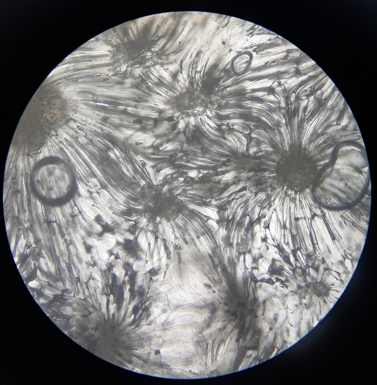
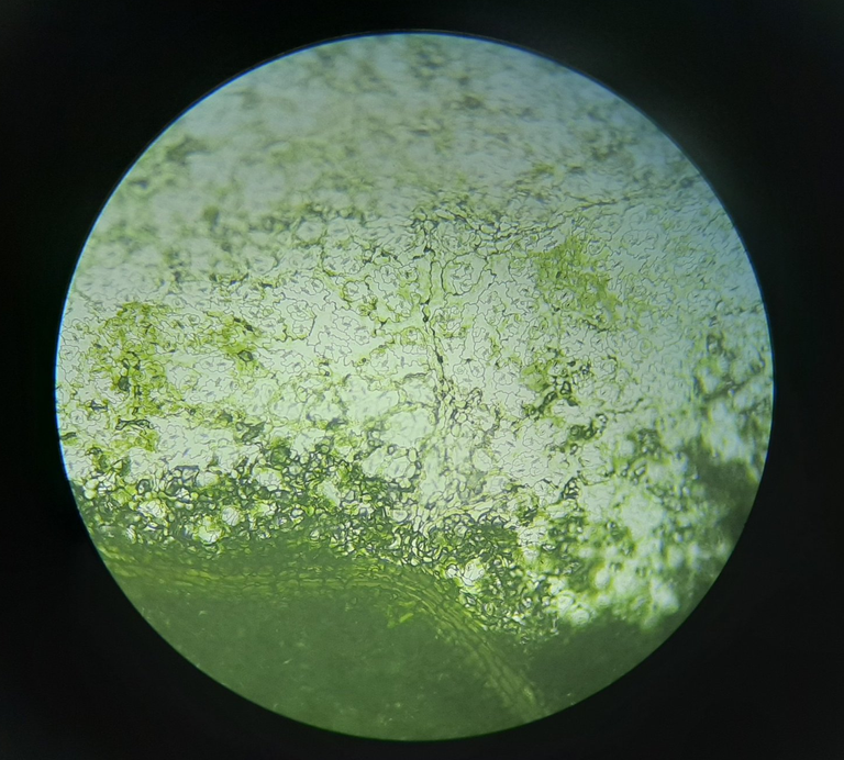
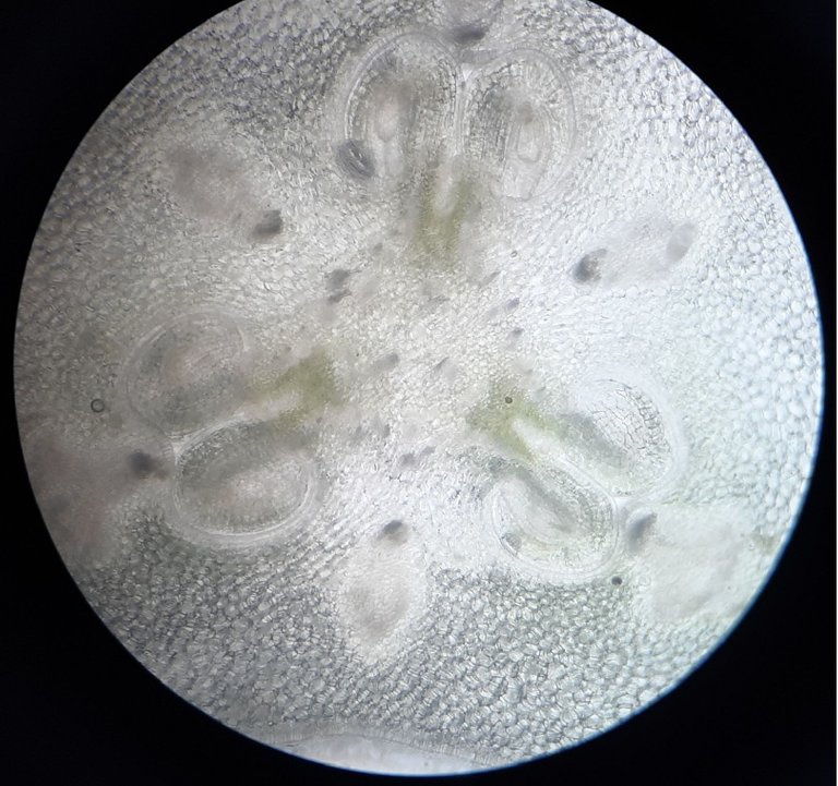
The special photo in today's lesson is to see the hard tissue cells of guava, the scientific name of guava is Psidium guajava, belonging to the family: Myrtaceae. The advantages of guava for health include Support healthy teeth and the immune system. beneficial to the brain,… Hard tissue cells of guavas are arranged in clusters and have the appearance of flowers. When viewed under a microscope, it is so amazing, and I am astonished by this particular picture.
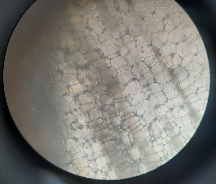
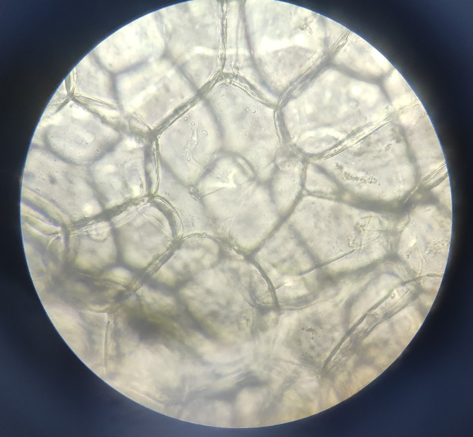
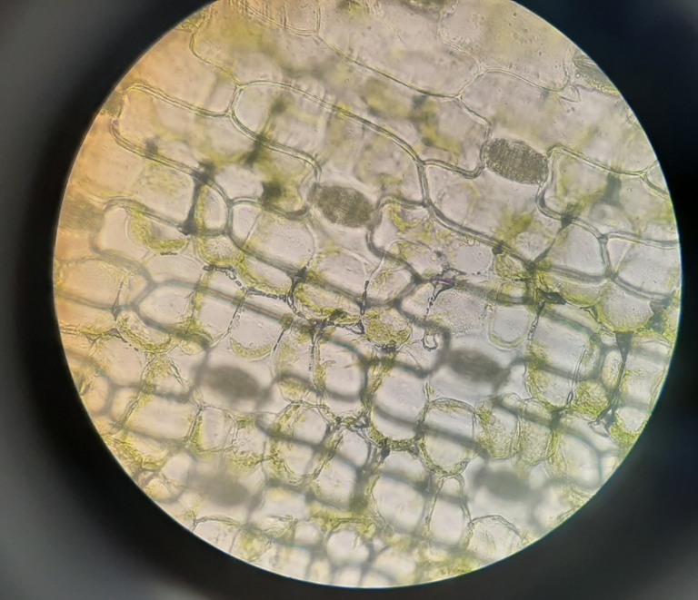
The onion's epidermal cells were the next thing I looked at. The genus Allium, which includes onions, is a member of the Alliaceae family of plants. Cells are arranged in a row, with a thin, colorless cell membranee. Onions have antioxidants, antimicrobial capabilities, blood sugar regulation, and heart health benefits.
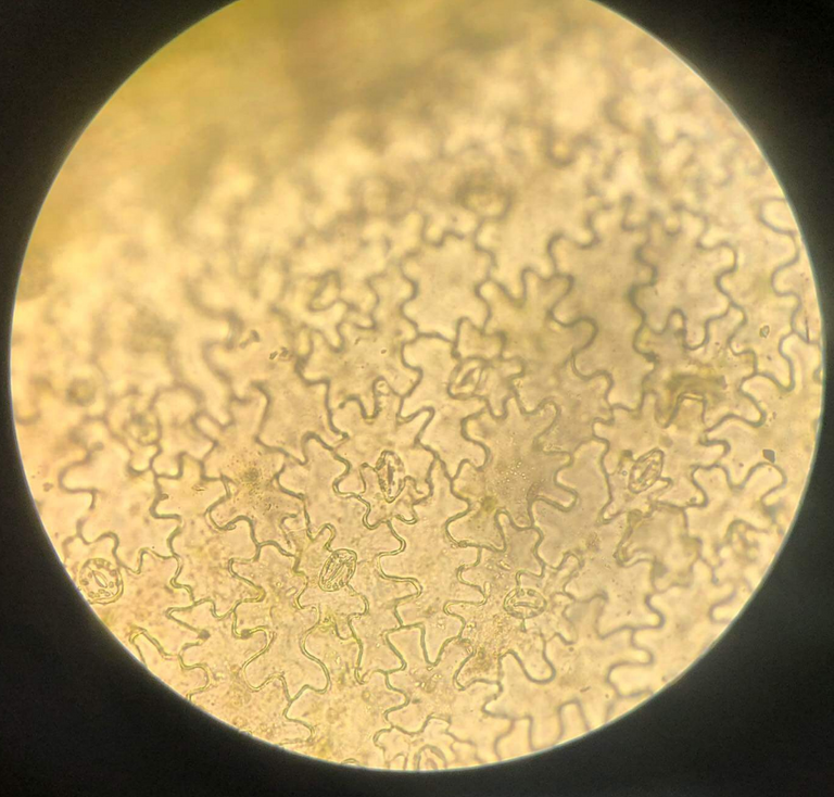
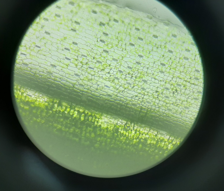
My next experiment involved perilla leaves. The scientific name for perilla, which is a member of the Lamiaceae family, is Perilla frutescens var. crispa. Perilla is used to treat heart disease, diabetes, asthma, and respiratory conditions. The stomatal cells in perilla are of the cross-celled type, and the stoma is encircled by two additional cells that are positioned perpendicular to the stoma's longitudinal axis.
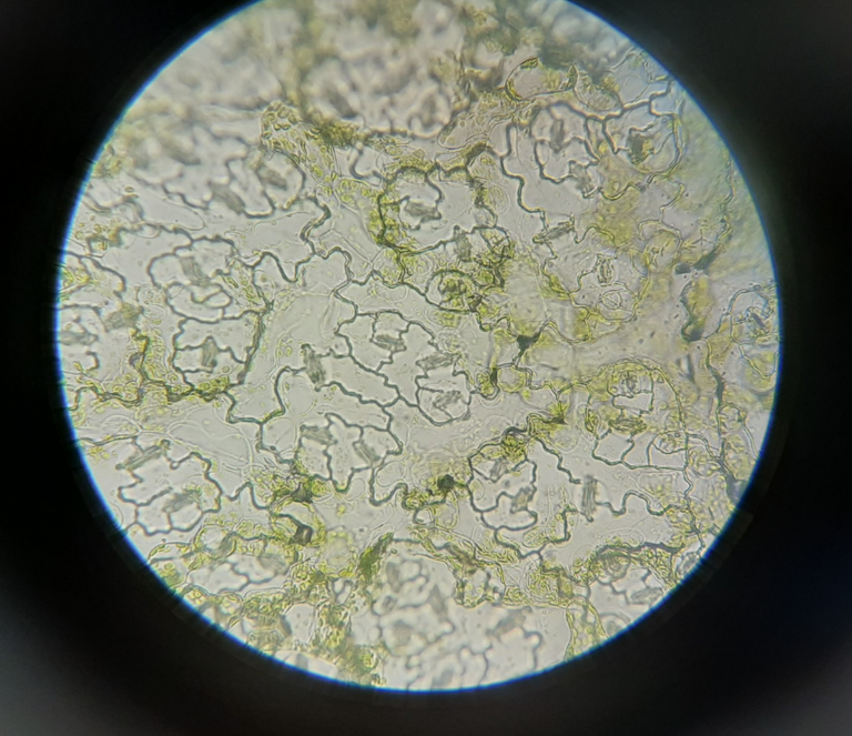
The leaves of Crinum latifolium L., a member of the Amaryllidaceae family, are the following item. The leaves of the crinum latifolium plant have the ability to ease bone pain. Parallel cells make up the stomatal cells.
Then comes the Brassicales family member, the mustard plant. Cruciferous vegetables have many advantages, including supporting liver function, protecting the heart,. Unequal-celled cellular structure, hree more cells surround the stoma, one of which is smaller than the other two.
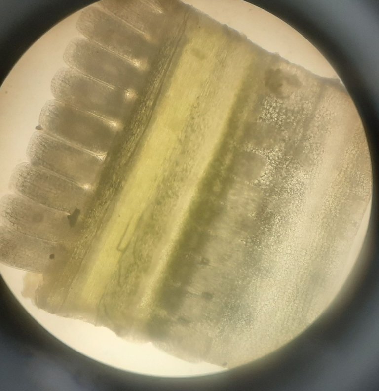
Coming to the second lesson, I discovered the design of the roots and leaves of two classes Magnoliidae and Liliopsida. I'll cut it into tiny slices with a cross cutter (or a vertical cut). Then dye the microsurgery and then water-rinse. I then move on to examine the microscope.
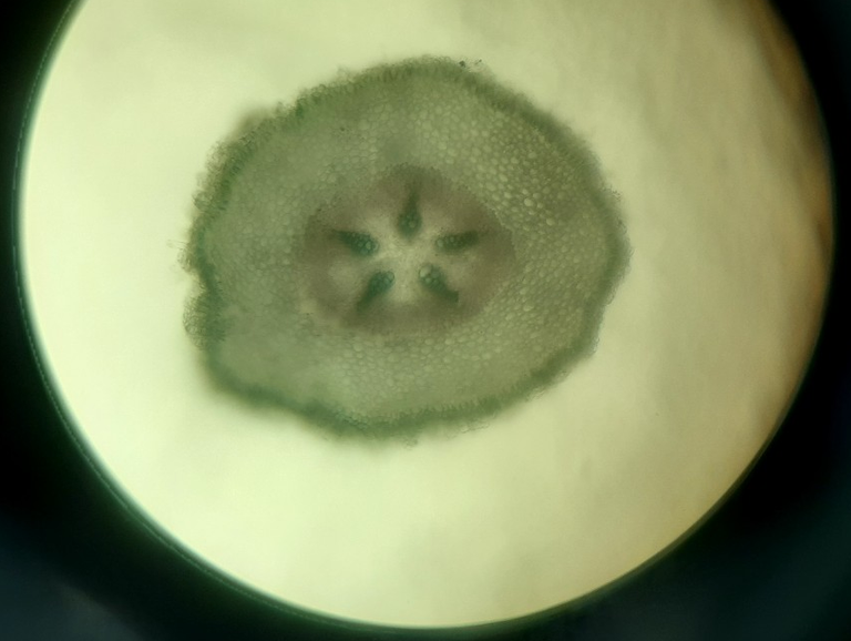
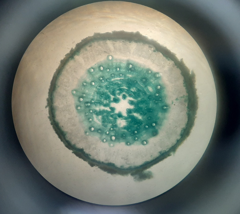
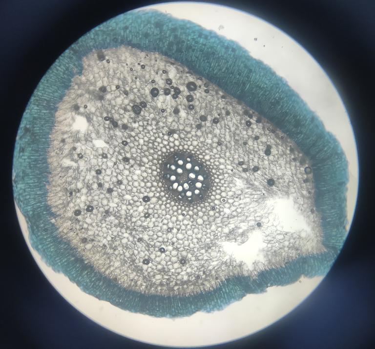
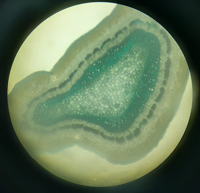
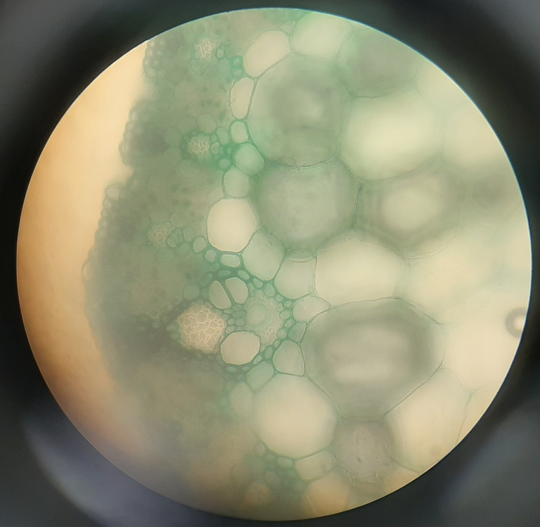
The leaves and roots of these 2 classes each have a different structure. They can be distinguished and recognized by this attribute as well.
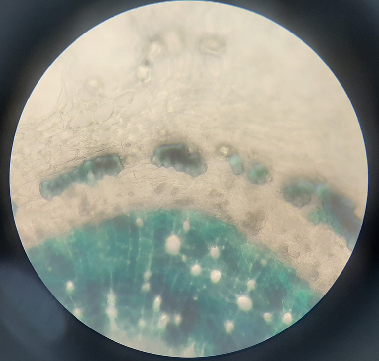
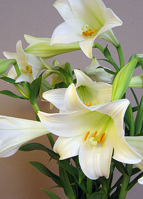
Next, I discovered more about lilies, commonly known as Lilium in science and a member of the Liliaceae family. When looking at leaves, petals, and ovary under a microscope, I learnt how to describe plants. Here are the lily images I captured.
These are really vivid photographs, I must say. These images provide new insights into plants' internal structures. The pictures you see above were all captured with my phone, and they must convince me that nature is magnificent. These images, in my opinion, are wonderful and have a unique beauty. This lecture taught me a lot about the native plants and their health advantages. It is very beneficial to my potential career. It was a wonderful learning experience, and I'm glad to share these experiences with everyone.
Thank you for taking the time to read my post. Have a nice day and get useful things from around. ❤️

Cultivating some plant joy 💚🌱 with a reshared!
Thank you very much for your comment. The plants is extremely rich and beautiful 😍
Wow!! I always wanted to do that!!! Never had a chance to get a good microscope to begin with but you basically shared something I always wanted to see.
Must have been pretty amazing~ how there is another universe in microscopic level.
Thanks for sharing. ;)
Must have been pretty amazing~ how there is another universe in microscopic level.
Thanks for sharing. ;)
Thanks for your comment. If you have a chance try to see everything under the microscope. There will be many surprises waiting for you 😁
Your major is cool! That's interesting to see things through a microscope view @strawberry89
Thank you so much for liking my post 😄
Congratulations @strawberry89! You have completed the following achievement on the Hive blockchain and have been rewarded with new badge(s):
Your next target is to reach 300 upvotes.
You can view your badges on your board and compare yourself to others in the Ranking
If you no longer want to receive notifications, reply to this comment with the word
STOPTo support your work, I also upvoted your post!
Check out the last post from @hivebuzz:
Support the HiveBuzz project. Vote for our proposal!
thanks for your support 😍
When I was in middle school, I also used to use a microscope to see all kinds of organisms in the water like Paramecium, Euglena, etc. This is very interesting.
Thank you for sharing, nice post.
Thank you so much for liking my post 😄
Oh, these are lovely! Please, experiment more in the lab and take more of these images! Perhaps it would lead to becoming an awesome NFT collection on the Tezos blockchain (which kind of brands itself as the blockchain for "green" NFTs).
Thank you for liking my post and pictures. I will update interesting things in upcoming posts. Please follow me 😘
Nice post
Thank you so much😄
Dear @strawberry89, we need your help!
The Hivebuzz proposal already got important support from the community. However, it lost its funding a few days ago and only needs a few more HP to get funded again.
May we ask you to support it so our team can continue its work this year?
You can do it on Peakd, ecency, or using HiveSigner.
https://peakd.com/me/proposals/199
Your support would be really appreciated.
Thank you!