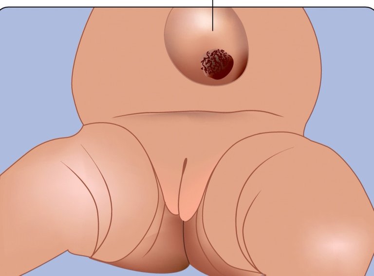
LAYERS OF THE ANTERIOR ABDOMINAL WALL The anterior abdominal wall is firm and elastic.
It consists of eight layers. From superficial to deep, these are: 1. Skin. 2. Superficial fascia. 3. External oblique muscle. 4. Internal oblique muscle. 5. Transversus abdominis muscle. 6. Fascia transversalis. 7. Extraperitoneal tissue. 8. Parietal layer of peritoneum.
The deep fascia is absent in the anterior abdominal wall to allow the bulging/distension of the abdominal wall as after taking meals, during pregnancy, etc. It is also absent in the penis, scrotum, and perineum.
Umbilicus 1. The umbilicus is the most obvious feature of the anterior abdominal wall. 2. It is in fact a normal puckered scar in the anterior abdominal wall representing the site of attachment of the umbilical cord in the fetus. Position The position of umbilicus is variable: 1. In adult, it lies at the level of intervertebral disc between L3 and L4 vertebrae. 2. In newborn, it is slightly at a lower level due to poorly developed pelvic region. 3. In old age, it comes down to lower level due to diminished tone of the abdominal muscles. Anatomical Significance 1. The level of umbilicus serves as water-shed line for venous and lymphatic drainage. The venous blood and lymph flow upward above the level of the umbilicus and downward below the level of the umbilicus. 2. It indicates the level of T10 dermatome, i.e., skin around the umbilicus is supplied by the 10th spinal segment. 3. It is one of the important sites of portocaval anastomosis.

Thanks for using eSteem!
Your post has been voted as a part of eSteem encouragement program. Keep up the good work!
Dear reader, Install Android, iOS Mobile app or Windows, Mac, Linux Surfer app, if you haven't already!
Learn more: https://esteem.app
Join our discord: https://discord.me/esteem
Great
Nice da
Hdjjdn
Useful one